COVID-19:富士フイルム、AIでコロナ診断支援:CT画像自動解析(動画):
COVID-19: Fujifilm, AI to support corona diagnosis: CT image automatic analysis:
COVID-19:Fujifilm,AI支持电晕诊断:CT图像自动分析
COVID-19:
2020年5月19日 17:45
富士フイルムホールディングス:
5月19日、人工知能(AI)を使って、新型コロナウイルス肺炎の診断を支援する技術を開発すると発表した。
CTで撮影した画像から病状を数値化し、医師が患者の経過や治療効果を判定するのを支援する。
このほど国内で共同研究を始め、2020年度中の製品化を目指す。
日本経済新聞
https://r.nikkei.com/article/DGXMZO59295700Z10C20A5XB0000?s=4
富士フイルム:新型コロナ肺炎の診断サポートAI開発:CT画像を自動解析
2020年5月19日
富士フイルム:
5月19日、新型コロナウイルス感染症による肺炎患者を受け入れている医療機関と共同で、AIを活用して患者の経過観察や治療効果の判断を支援する技術を開発すると発表した。
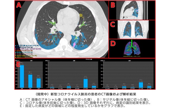
胸部のCT画像を自動解析し、肺炎による異常を識別するという。
同社が2018年から京都大学と共同研究していた技術を応用する。
- 肺を12の領域に分け、
- 気管支や肺の血管などに
- 異常部分の大きさや割合などを判定。
- 医師はその情報を基に、病状の確認や治療の効果を判断する。
京都大学:共同研究
新型コロナウイルス感染症による肺炎患者を治療する際、医師は刻々と変化する病状をCT画像などから目視で確認する。
しかし、CT画像は患者1人当たり数百枚にも及ぶため、医師の負担になっている。
同社は薬の効果計測にも使えるとして、新型コロナウイルス感染症の治療薬の開発や評価にも貢献したい考え。
ITmedia NEWS
https://www.itmedia.co.jp/news/spv/2005/19/news105.html
Fujifilm to develop AI-based technology to aid COVID-19-
induced pneumonia diagnosis and assess the effectiveness of treatments | Fujifilm Global
TOKYO, May 19, 2020 —
FUJIFILM Corporation (President: Kenji Sukeno) is commencing a research study to develop Artificial Intelligence (AI)-based technology to aid in the diagnosis and treatment assessment of patients with COVID-19-induced pneumonia.
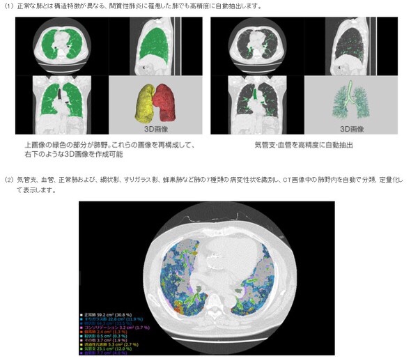
The technology for quantifying the lesions of interstitial pneumonia*, co-developed with Kyoto University (the Department of Respiratory Medicine, Graduate School of Medicine, Professor Toyohiro Hirai), will be applied to the project.
The company will now embark on a joint research study with local medical institutions treating COVID-19 patients, starting with the Kanagawa Cardiovascular and Respiratory Center (Yokohama, Japan).
The spread of the novel coronavirus, which causes COVID-19, has emerged as a serious issue around the world.
The world has yet to see clear judging criteria for determining the effectiveness of various treatment options, currently explored by doctors. In order to confirm the progression of pneumonia and the effectiveness of treatments,
doctors need to examine hundreds of chest CT images for each patient to visually check the characteristics of ever-changing lesions and it puts a serious strain on specialists.
There are expert opinions that COVID-19-induced pneumonia presents similarly to interstitial pneumonia in diagnostic images and has diverse variations in lesion patterns.
Fujifilm’s CT quantification technology for interstitial pneumonia is powered by an AI-based software
that examines CT images to identify
- bronchi,
- blood vessels and
- normal lungs in lung field**
as well as seven types of lesions such as
- reticular opacities,
- ground-glass opacities and
- honeycomb lungs***,
and automatically carries out categorization and measurement to quantify lesions of interstitial pneumonia.
It also divides the lung field into 12 zones*4
and shows the volume and ratio of lesions for each of the zones so that clinicians can examine the distribution and progression of lesions within the lung field in details.