COVID-19:Fujifilm,AI支持电晕诊断:CT图像自动分析
COVID-19:
2020年5月19日17:45
富士胶片控股公司:
5月19日,宣布将开发利用人工智能(AI)支持新冠状病毒性肺炎诊断的技术。
通过从CT拍摄的图像对医疗状况进行数字化,它可以帮助医生确定患者的病情和治疗效果。
我们已经在日本开始了联合研究,并致力于在2020财年末实现商业化。
日本经济新闻
https://r.nikkei.com/article/DGXMZO59295700Z10C20A5XB0000?s=4
FUJIFILM:AI支持诊断新的日冕性肺炎:CT图像的自动分析
2020年5月19日
FUJIFILM:
5月19日,宣布将与AI共同与一家接受新型冠状病毒感染的肺炎患者的医疗机构合作,共同开发一种技术,以支持患者的随访和判断疗效。
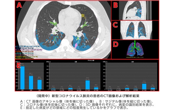
据说会自动分析胸部的CT图像,以识别由于肺炎引起的异常。
自2018年以来,应用公司与京都大学进行联合研究的技术。
将肺分为12个区域,
用于肺的支气管和血管
确定异常零件的大小和比例。
根据该信息,医生确认病情并确定治疗效果。
京都大学:共同研究
当治疗由于新的冠状病毒感染引起的肺炎时,医生会从CT图像上目视确认不断变化的医疗状况。
但是,每位患者有数百张CT图像,这给医生带来了负担。
该公司希望为新的冠状病毒感染的治疗剂的开发和评估做出贡献,并称其可用于测量药物的作用。
IT媒体新闻
https://www.itmedia.co.jp/news/spv/2005/19/news105.html
Fujifilm to develop AI-based technology to aid COVID-19-
induced pneumonia diagnosis and assess the effectiveness of treatments | Fujifilm Global
TOKYO, May 19, 2020 —
FUJIFILM Corporation (President: Kenji Sukeno) is commencing a research study to develop Artificial Intelligence (AI)-based technology to aid in the diagnosis and treatment assessment of patients with COVID-19-induced pneumonia.
The technology for quantifying the lesions of interstitial pneumonia*, co-developed with Kyoto University (the Department of Respiratory Medicine, Graduate School of Medicine, Professor Toyohiro Hirai), will be applied to the project.
The company will now embark on a joint research study with local medical institutions treating COVID-19 patients, starting with the Kanagawa Cardiovascular and Respiratory Center (Yokohama, Japan).
The spread of the novel coronavirus, which causes COVID-19, has emerged as a serious issue around the world.
The world has yet to see clear judging criteria for determining the effectiveness of various treatment options, currently explored by doctors. In order to confirm the progression of pneumonia and the effectiveness of treatments,
doctors need to examine hundreds of chest CT images for each patient to visually check the characteristics of ever-changing lesions and it puts a serious strain on specialists.
There are expert opinions that COVID-19-induced pneumonia presents similarly to interstitial pneumonia in diagnostic images and has diverse variations in lesion patterns.
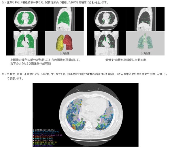
Fujifilm’s CT quantification technology for interstitial pneumonia is powered by an AI-based software
that examines CT images to identify
- bronchi,
- blood vessels and
- normal lungs in lung field**
as well as seven types of lesions such as
- reticular opacities,
- ground-glass opacities and
- honeycomb lungs***,
and automatically carries out categorization and measurement to quantify lesions of interstitial pneumonia.
It also divides the lung field into 12 zones*4
and shows the volume and ratio of lesions for each of the zones so that clinicians can examine the distribution and progression of lesions within the lung field in details.