東大/阪大:定量位相・分子振動顕微鏡;細胞形態/分子分布、同時観察:
Unprecedented 3D images of live cells plus details of molecules:
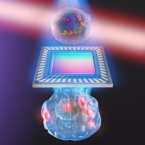
前所未有的活细胞3D图像以及内部分子的详细信息新成像方法的艺术表现形式
~標識不要な定量位相顕微鏡と分子振動顕微鏡のコラボレーション~
東大/阪大:
蛍光標識なしに、「細胞の高解像形態画像に、生体分子の分布を示せる光学顕微鏡」を、開発した。
- 赤外光で細胞内分子に対し
- 固有の振動を生じさせ、
- 分子振動に伴う温度上昇による
- 屈折率の変化を、計測する原理。
今回の光学顕微鏡:
「生体分子の分布を観察し、細胞形態と分子分布の情報を同時に取得すること」に、成功した。
「生きたままの細胞観察が必要となる再生医療や生物学研究への幅広い利用」が期待される。
従来の顕微鏡:蛍光標識
生命科学/医療分野では、細胞や細胞内小器官の形態や細胞構成分子の分布を、複数の顕微鏡を用いて観察します。
その中でも、「生体分子を蛍光標識し、細胞内の動態を顕微鏡で観察する手法」が、広く利用されています。
蛍光物質を結合させることで、生体分子の生理活性を阻害します。
また、標識をつけた分子を細胞内に入れるため、細胞の一部を壊します。
今回の顕微鏡:非標識(ラベルフリー):
そこで、生体分子を非標識(ラベルフリー)にする顕微鏡が待たれていました。
つまり、生きたままの状態で観察できる顕微鏡(ラベルフリー顕微鏡)の開発です。
非標識(ラベルフリー):2種類
従来のラベルフリー顕微鏡には、
- 細胞の形態を詳細に測る定量位相顕微鏡、
- 生体分子の分布を測る分子振動顕微鏡、
- 2種類があります。
しかし、それらの顕微鏡では、「細胞の形態」もしくは「分子の分布」のいずれか一方の計測しかできませんでした。
東京大学/大阪大学:
- 定量位相顕微鏡技術:可視光で高解像形態画像を計測、
- 分子振動分光技術:赤外光で分子振動を計測、
- 融合した、新しい赤外フォトサーマル定量位相顕微鏡の開発に成功しました。
この新しい顕微鏡技術:
- 細胞を破壊することなく、
- 細胞の詳細な形態情報と
- 分子分布情報の同時計測を
- 実現できます。
医療や生物学における新たな細胞計測ツールとして利用されることが期待されます。
本研究成果は、2020年4月20日(米国東部夏時間)に「Optica」にオンライン掲載されます。
https://www.jst.go.jp/pr/announce/20200420-2/index.html
https://www.jst.go.jp/pr/announce/20200420-2/pdf/20200420-2.pdf
Unprecedented 3D images of live cells plus details of molecules inside An artistic representation of the new imaging method
called biochemical quantitative phase imaging with midinfrared photothermal effect.
© 2020 s-graphics.co.jp, CC BY-NC-ND.
- Collaborators at Osaka University,
- other departments at the University of Tokyo and
- the Japan Science and Technology Agency also contributed to this research.
The insides of living cells
can be seen in their natural state in greater detail than ever before using a new technique developed by researchers in Japan.
This advance should help reveal the complex and fragile biological interactions of medical mysteries, like how stem cells develop or how to deliver drugs more effectively.
“Our system is based on a simple concept, which is one of its advantages,”
said Associate Professor Takuro Ideguchi from the University of Tokyo Research Institute for Photon Science and Laser Technology.
The results of Ideguchi’s team were published recently in Optica, the Optical Society’s research journal.
The new method also
has the advantages of not needing to kill the cells, damage them with intense light, or artificially attach fluorescent tags to specific molecules.
The technique
combines two pre-existing microscopy tools and uses them simultaneously. The combination of these tools can be thought of simply as like a coloring book.
“We gather the black-and-white outline of the cell and we virtually color in the details about where different types of molecules are located,” said Ideguchi.
Quantitative phase microscopy
gathers information about the black-and-white outline of the cell using pulses of light and measuring the shift in the light waves after they pass through a sample.
This information is used to reconstruct a 3D image of the major structures inside the cell.
Molecular vibrational imaging
provides the virtual color using pulses of midinfrared light to add energy to specific types of molecules.
That extra energy causes the molecules to vibrate, which heats up their local surroundings. Researchers can choose to raise the temperature of specific types of chemical bonds by using different wavelengths of midinfrared light.
Researchers
take a quantitative phase microscopy image of the cell with the midinfrared light turned off and an image with it turned on.
The difference between those two images then reveals both the outline of major structures inside the cell and the exact locations of the type of molecule that was targeted by the infrared light.
Researchers
refer to their new combined imaging method as biochemical quantitative phase imaging with midinfrared photothermal effect.
“We were impressed when we first observed the molecular vibrational signature characteristic of proteins, and we were further excited when this protein-specific signal appeared in the same location as the nucleolus, an intracellular structure where high amounts of proteins would be expected,” said Ideguchi.
Ideguchi’s team
hopes their technique might allow researchers to determine the distribution of fundamental types of molecules inside single cells.
The quantitative phase microscopy outline of major structures
could be virtually colored in using different wavelengths of light to specifically target proteins, lipids (fats) or nucleic acids (DNA, RNA).
Currently,
capturing one complete image can take 50 seconds or longer.
Researchers are confident that they can speed up the process
with simple improvements to their tools, including a higher-powered light source and a more sensitive camera.
The University of Tokyo