iPS cells: creating the base of the skeleton!
ーToward development of treatments for spinal diseasesー
Kyoto University:
Using iPS cells, inside a fertilized egg (embryo),
We created a model of a somite, a mass of cells that form the basis of the skeleton and muscles.
On December 21, it was announced in the British scientific journal Nature.
Studies with human embryos:
Research using human embryos is strictly restricted from the viewpoint of respect for human life.
By this method,
without using human embryos.
elucidation of the process by which an embryo develops into a fetus,
This will lead to the development of treatments for congenital spinal disorders.
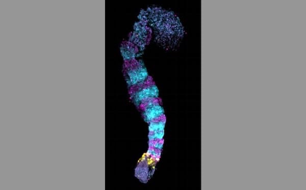
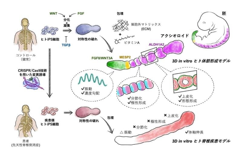
In vitro somite modeling:
A mass of human iPS cells was cultured three-dimensionally to create a somite model in vitro.
Similar in shape to human embryos.
It was also confirmed that similar genes were expressed.
Reproducing pathology with iPS cells:
“iPS cells with genetic mutations found in patients with congenital spinal disease”
This model was created to reproduce the pathology of the disease.
The research team says, “We would like to understand the mechanism of biogenesis.”
Nihon Keizai Shimbun
https://www.nikkei.com/article/DGXZQOUF21ANT0R21C22A2000000/
Cellules iPS : créer la base du squelette !
ーVers le développement de traitements pour les maladies de la colonne vertébraleー
Université de Kyôto :
À l’aide de cellules iPS, à l’intérieur d’un œuf fécondé (embryon),
Nous avons créé un modèle de somite, une masse de cellules qui forment la base du squelette et des muscles.
Le 21 décembre, il a été annoncé dans la revue scientifique britannique Nature.
Études avec des embryons humains :
La recherche utilisant des embryons humains est strictement encadrée du point de vue du respect de la vie humaine.
Par cette méthode,
sans utiliser d’embryons humains.
élucidation du processus par lequel un embryon se transforme en fœtus,
Cela conduira au développement de traitements pour les troubles rachidiens congénitaux.
Modélisation des somites in vitro :
Une masse de cellules iPS humaines a été cultivée en trois dimensions pour créer un modèle somite in vitro.
De forme similaire à celle des embryons humains.
Il a également été confirmé que des gènes similaires étaient exprimés.
Reproduire la pathologie avec des cellules iPS :
“Cellules iPS avec des mutations génétiques trouvées chez des patients atteints de maladie congénitale de la colonne vertébrale”
Ce modèle a été créé pour reproduire la pathologie de la maladie.
L’équipe de recherche déclare : « Nous aimerions comprendre le mécanisme de la biogenèse.
Nihon Keizai Shimbun
iPS-Zellen: Erstellen der Basis des Skeletts!
ーEntwicklung von Behandlungen für Wirbelsäulenerkrankungenー
Universität Kyoto:
Unter Verwendung von iPS-Zellen in einem befruchteten Ei (Embryo)
Wir haben ein Modell eines Somiten erstellt, einer Masse von Zellen, die die Basis des Skeletts und der Muskeln bilden.
Am 21. Dezember wurde es im britischen Wissenschaftsjournal Nature angekündigt.
Studien mit menschlichen Embryonen:
Forschung mit menschlichen Embryonen ist aus Sicht der Achtung des menschlichen Lebens strengstens untersagt.
Durch diese Methode
ohne Verwendung menschlicher Embryonen.
Aufklärung des Prozesses, durch den sich ein Embryo zu einem Fötus entwickelt,
Dies wird zur Entwicklung von Behandlungen für angeborene Wirbelsäulenerkrankungen führen.
In-vitro-Somit-Modellierung:
Eine Masse menschlicher iPS-Zellen wurde dreidimensional kultiviert, um ein Somitenmodell in vitro zu erstellen.
Ähnlich in der Form wie menschliche Embryonen.
Es wurde auch bestätigt, dass ähnliche Gene exprimiert wurden.
Reproduktion von Pathologien mit iPS-Zellen:
“iPS-Zellen mit genetischen Mutationen bei Patienten mit angeborener Wirbelsäulenerkrankung gefunden”
Dieses Modell wurde erstellt, um die Pathologie der Krankheit zu reproduzieren.
„Wir möchten den Mechanismus der Biogenese verstehen“, sagt das Forschungsteam.
Nihon Keizai Shimbun
Reconstituting human somitogenesis in vitro
Nature
Abstract
The segmented body plan of vertebrates is established during somitogenesis, a well-studied process in model organisms,
but remains largely elusive in humans due to ethical and technical limitations.
Despite recent advances with pluripotent stem cell (PSC)-based approaches1-5,
models that robustly recapitulate human somitogenesis in both space and time are still largely missing.
Here,
we introduce a PSC-derived mesoderm-based 3D model of human segmentation and somitogenesis, which we termed ‘axioloid’,
that captures accurately the oscillatory dynamics of the segmentation clock and the morphological and molecular characteristics of sequential somite formation in vitro.
Axioloids show proper rostrocaudal patterning of forming segments
and robust anterior-posterior FGF/WNT signaling gradients and retinoic acid (RA) signaling components.
We identify an unexpected critical role of RA signaling in the stabilization of forming segments, indicating distinct,
but also synergistic effects of RA and extracellular matrix (ECM) on the formation and epithelialization of somites.
Importantly,
comparative analysis demonstrates striking similarities of axioloids to the human embryo, further validated by the presence of a HOX code in axioloids.
Lastly,
we demonstrate the utility of axioloids to study the pathogenesis of human congenital spine diseases,
by using patient-like iPSCs with mutations in HES7and MESP2.
These results suggest that axioloids represent a promising novel platform to study axial development and disease in humans.