The University of Tokyo / JEOL: Direct observation of magnetic field at atomic level:
-Development of high-performance electron microscope MARS-
University of Tokyo
Electron microscope / JEOL
The research team has developed a high-performance electron microscope “Atomic Resolution Magnetic Field Free Electron Microscope (MARS)”.
We succeeded in directly observing the origin of magnets and magnetic fields at the atomic level for the first time in the world.
It was announced in the electronic version of the English scientific journal Nature on February 9.
Atomic Resolution Magnetic Field Free Electron Microscope (MARS):
This observation technique
Magnets and magnetic semiconductors,
Magnetic memory, etc.
Useful for a wide range of research and development.
Professor Naoya Shibata of the University of Tokyo:
It increases the storage capacity of magnetic memory and makes EV magnets less likely to deteriorate.
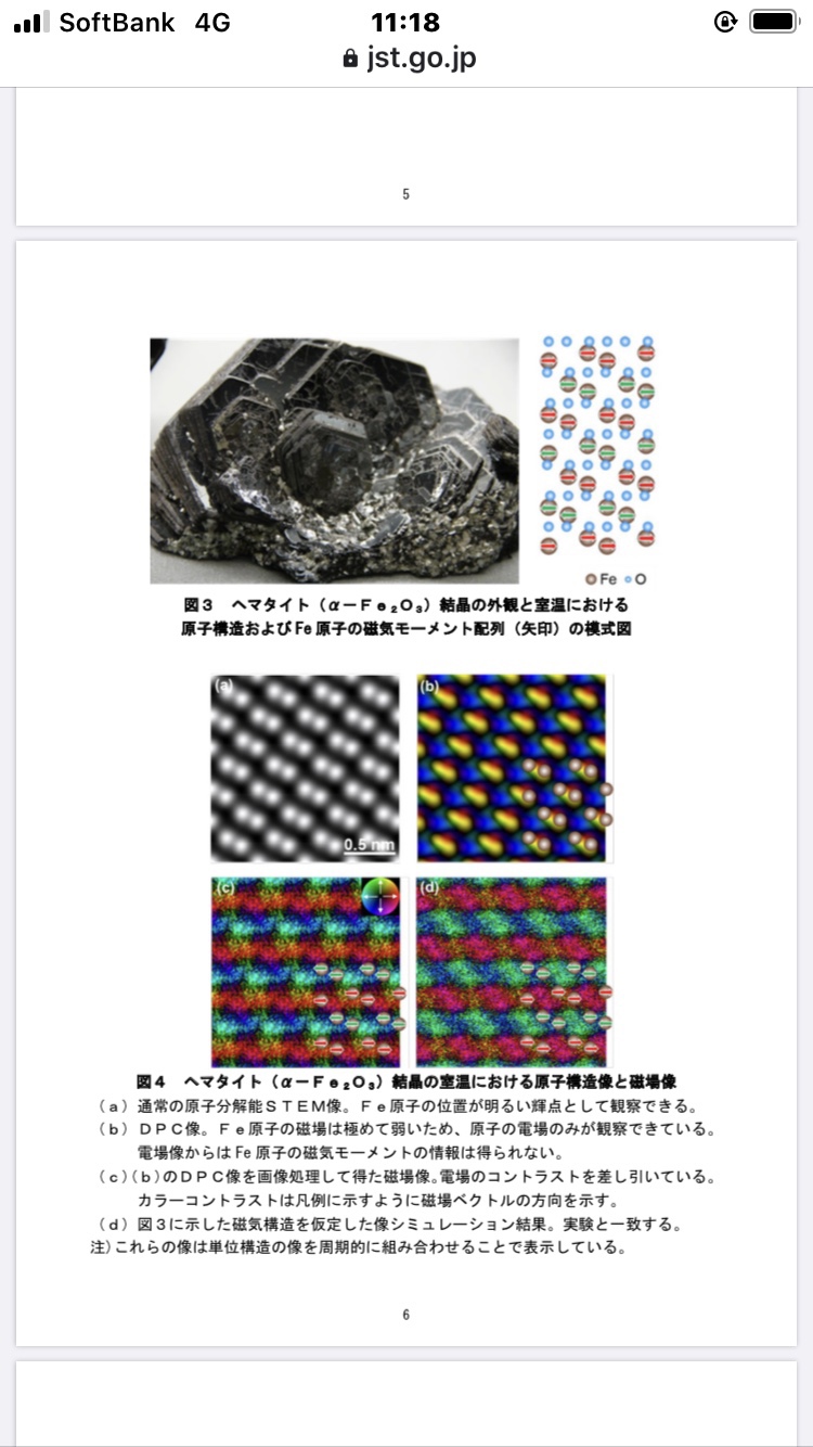
Direct observation of atomic level magnetic field:
The object of this observation is a kind of iron ore,
Hematite An iron atom in a crystal.
In this crystal, layers of iron atoms and layers of oxygen atoms are alternately stacked.
When cooled significantly, the direction of the magnetic field was rotated 90 degrees and became vertical, which was in agreement with the result of the theoretical calculation.
Conventional problems:
Place the electron microscope in a strong magnetic field and place the sample you want to observe in a strong magnetic field.
When an electron beam is applied, the magnetic field acts as a magnifying lens.
However, if the sample itself is a magnetic material, it may not be observed well.
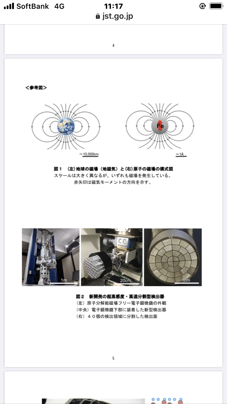
Developed new MARS:
In 2019, the magnetic field acting as a magnifying lens was set to two stages, upper and lower.
We have developed “MARS that cancels each other’s magnetic fields only in the part where the sample is placed”.
afterwards,
The electric and magnetic fields of the sample atom were separated by a filter.
“Ultra-sensitive detector that captures weak signal of magnetic field” was combined.
Current affairs dot com
https://www.jiji.com/jc/article?k=2022021000046&g=soc
Joint presentation: Capturing the origin of magnetic force with the world’s first atomic-resolution electron microscope-accelerating research and development of cutting-edge materials such as magnets, semiconductors, and quantum technology-
https://www.jst.go.jp/pr/announce/20220210/index.html
Real-space visualization of intrinsic magnetic fields of an antiferromagnet
Nature
Abstract
Characterizing magnetic structures down to atomic dimensions
is central to the design and control of nanoscale magnetism in materials and devices.
However,
real-space visualization of magnetic fields at such dimensions has been extremely challenging.In recent years,
atomic-resolution differential phase contrast scanning transmission electron microscopy (DPC STEM)1has enabled direct imaging of electric field distribution even inside single atoms2.
Here we show
real-space visualization of magnetic field distribution inside antiferromagnetic haematite (α-Fe2O3) using atomic-resolution DPC STEM in a magnetic-field-free environment3.After removing the phase-shift component due to atomic electric fields and improving the signal-to-noise ratio by unit-cell averaging,
real-space visualization of the intrinsic magnetic fields in α-Fe2O3 is realized.
These results open a new possibility for real-space characterization of many magnetic structures.