Figure 1 Three-dimensional fluorescence imaging by nonlinear optical phenomenon
Fluorescence generated by a non-linear optical phenomenon is detected by using a near-infrared laser beam having high tissue permeability. Therefore, even a living tissue that has not been fixed or stained can be three-dimensionally observed up to the deep part.
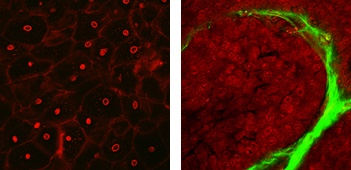
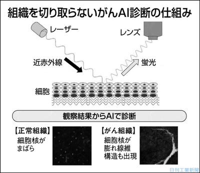
Figure 2 Imaging images of normal human cervical tissue and cervical cancer
(Left): Imaging image of normal tissue. Cell nuclei (red) are sparse and no fibrous structure (green) can be recognized around the cells.
(Right): Imaging image of cervical cancer. The nuclei (red) of cancer cells are swollen and dense. A fibrous structure (green) appears around the cells.
Osaka Univ.: Cancer, immediate diagnosis without cutting: near-infrared laser/biological imaging
2020/7/23 23:00
Osaka University, Kyushu University, Nikon: Research Team:
Traditional cancer final diagnosis:
Traditionally, a pathologist has cut out a suspected part of cancer and observed and diagnosed it with a microscope.
However, it requires a formalin fixation procedure to make observation specimens, which is time-consuming.
In addition, there were “issues such as the diagnosis cannot be confirmed when the number of samples is small”.
This research team:
Utilizing the technology of “living body imaging that captures the emitted light by applying a near infrared laser to the tissue”.
Proved that it can be observed in 3D without cutting or fixing cancer tissue.
Asahi Shimbun Digital
https://www.asahi.com/sp/articles/ASN7P622DN7LPLBJ002.html?ref=amp_login
Immediately diagnose cancer on the spot without cutting “light biopsy”
-Stereoscopic observation without damage to tissue by imaging, AI automatically diagnoses-
July 23, 2020 Press Release
Osaka University
Kyushu University
Nikon Corporation
Japan Medical Research and Development Organization
Points of research results:
Application of biological imaging, observation technology that allows human tissue to be visualized in 3D as it is without being cut from the human body and stained
By using image analysis by AI together, cervical cancer can be diagnosed “without damage”, “quickly” and “quantitatively” compared to conventional diagnostic methods.
Providing cancer tissue diagnosis through IoT to regions with few medical professionals including pathologists in developing countries
Overview
The research group
We have developed a method that enables real-time three-dimensional observation of the living tissue of the cervix without formalin fixation or staining.
By using this observation method, which does not require tissue cutting, and image analysis by AI together, cervical cancer and its ultra-early lesions can be classified quantitatively without damage.
The results of this research will be published in the online version of the Cancer Research journal “Cancer Research” at 23 o’clock (JST) on July 23, 2020.
Japan Medical Research and Development Organization