Novel Coronavirus: Fine Visualization!
– Kao prepares stained virus particles –
Kao:
-VHH antibody that binds to the novel coronavirus-
Using “a fusion protein linked with a high-intensity fluorescent protein”,
We have been studying “application to diagnosis of new coronavirus”.
Prepare stained virus particles:
We have developed a simple visualization technology for viruses in the environment.
Novel coronavirus:
Treat with paraformaldehyde.
Viral RNA is stained after infectivity is eliminated.
Add SYTO82, a KikG-fused VHH antibody that binds to the S protein.
Prepare stained virus particles.
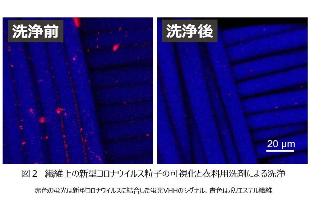
Virus attached to textiles:
The prepared virus particles were dropped onto a polyester cloth to attach the virus to the fiber.
Observed under a fluorescence microscope:
Observed with a fluorescence microscope.
on the fiber
Virus-derived fluorescence (SYTO82) and KikG-fused VHH antibody-derived fluorescence
detected at the same position.
Visualization of virus particles:
This result is
Virus particles attached to the fiber are brightly fluorescent from the KikG-fused VHH antibody,
It shows that it is visualized.
Wash with laundry detergent:
I washed the virus-attached cloth with laundry detergent.
Fluorescence derived from KikG-VHH fusion protein
It is confirmed that it decreases significantly from the fiber.
This test:
“Possibility of visualizing viruses even on textiles” was shown.
Until now, it could only be confirmed with an electron microscope.
Viral particles in the environment:
“High-brightness/fluorescent protein” and “VHH antibody with high binding ability”
By binding, virus particles can be easily observed.
Forbes JAPAN Official Site
https://forbesjapan.com/articles/detail
Nouveau coronavirus : belle visualisation !
– Kao prépare des particules virales colorées –
Kaô :
-Anticorps VHH qui se lie au nouveau coronavirus-
En utilisant “une protéine de fusion liée à une protéine fluorescente de haute intensité”,
Nous étudions “l’application au diagnostic du nouveau coronavirus”.
Préparez les particules virales colorées :
Nous avons développé une technologie simple de visualisation des virus dans l’environnement.
Coronavirus nouveau:
Traiter avec du paraformaldéhyde.
L’ARN viral est coloré après élimination de l’infectiosité.
Ajouter SYTO82, un anticorps VHH fusionné avec KikG qui se lie à la protéine S.
Préparer les particules virales colorées.
Virus attaché aux textiles :
Les particules virales préparées ont été déposées sur un tissu en polyester pour attacher le virus à la fibre.
Observé au microscope à fluorescence :
Observé au microscope à fluorescence.
sur la fibre
Fluorescence dérivée du virus (SYTO82) et fluorescence dérivée de l’anticorps VHH fusionné avec KikG
détecté au même endroit.
Visualisation des particules virales :
Ce résultat est
Les particules virales attachées à la fibre sont brillamment fluorescentes à partir de l’anticorps VHH fusionné avec KikG,
Cela montre qu’il est visualisé.
Laver avec un détergent à lessive :
J’ai lavé le chiffon infecté par le virus avec un détergent à lessive.
Fluorescence dérivée de la protéine de fusion KikG-VHH
Il est confirmé qu’elle diminue significativement à partir de la fibre.
Ce test:
“Possibilité de visualiser des virus même sur des textiles” a été démontrée.
Jusqu’à présent, cela ne pouvait être confirmé qu’au microscope électronique.
Particules virales dans l’environnement :
“Protéine à haute luminosité/fluorescente” et “anticorps VHH avec une capacité de liaison élevée”
En se liant, les particules virales peuvent être facilement observées.
Forbes JAPON Site officiel
Novel Coronavirus: Feine Visualisierung!
– Kao bereitet gefärbte Viruspartikel vor –
Kao:
-VHH-Antikörper, der an das neuartige Coronavirus bindet-
Verwendung “eines Fusionsproteins, das mit einem hochintensiven fluoreszierenden Protein verbunden ist”,
Wir haben die „Anwendung zur Diagnose des neuen Coronavirus“ untersucht.
Bereiten Sie gefärbte Viruspartikel vor:
Wir haben eine einfache Visualisierungstechnologie für Viren in der Umgebung entwickelt.
Neuartiges Coronavirus:
Mit Paraformaldehyd behandeln.
Virale RNA wird gefärbt, nachdem die Infektiosität eliminiert wurde.
Fügen Sie SYTO82 hinzu, einen KikG-fusionierten VHH-Antikörper, der an das S-Protein bindet.
Bereiten Sie gefärbte Viruspartikel vor.
An Textilien haftende Viren:
Die hergestellten Viruspartikel wurden auf ein Polyestertuch getropft, um das Virus an der Faser zu befestigen.
Unter einem Fluoreszenzmikroskop beobachtet:
Beobachtet mit einem Fluoreszenzmikroskop.
auf der Faser
Virus-abgeleitete Fluoreszenz (SYTO82) und KikG-fusionierte VHH-Antikörper-abgeleitete Fluoreszenz
an der gleichen Stelle erkannt.
Visualisierung von Viruspartikeln:
Dieses Ergebnis ist
An der Faser anhaftende Viruspartikel fluoreszieren hell vom KikG-fusionierten VHH-Antikörper,
Es zeigt, dass es visualisiert wird.
Mit Waschmittel waschen:
Ich habe das virusbehaftete Tuch mit Waschmittel gewaschen.
Fluoreszenz stammt von KikG-VHH-Fusionsprotein
Es wird bestätigt, dass es von der Faser signifikant abnimmt.
Dieser Test:
„Möglichkeit Viren auch auf Textilien zu visualisieren“ wurde gezeigt.
Bisher konnte es nur mit einem Elektronenmikroskop bestätigt werden.
Viruspartikel in der Umwelt:
„High-Brightness/Fluoreszenz-Protein“ und „VHH-Antikörper mit hoher Bindungsfähigkeit“
Durch die Bindung können Viruspartikel leicht beobachtet werden.
Offizielle Seite von Forbes JAPAN
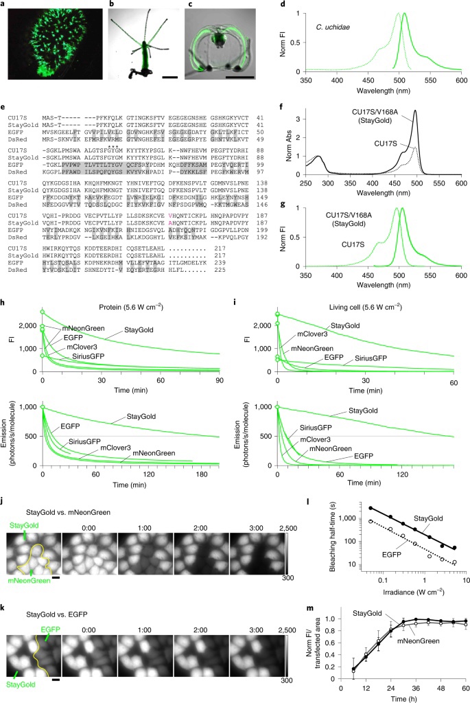
Kao | A Highly Photostable Fluorescent Protein
– Technology That Allows For Quantitative Observation of Cellular Microstructures and Viruses –
A joint research group consisting of researchers from
the Laboratory for Cell Function Dynamics of the RIKEN Center for Brain Science
the Biotechnological Optics Research Team of the RIKEN Center for Advanced Photonics;
the Asamushi Research Center for Marine Biology, Graduate School of Life Sciences, Tohoku University;
the Ōmura Satoshi Memorial Institute, Kitasato University;
the Safety Science Research Laboratories,
Kao Corporation,
promotes sustainable bioimaging by utilizing a jellyfish-derived green fluorescent protein StayGold, which is bright and exhibits minimal fading.
StayGold improves
spatiotemporal resolution and dramatically extends the observation period.
This finding offers
a new and powerful tool for the bioimaging community,
in which most experimenters encounter the challenging issue of photobleaching[1] of fluorescent proteins.
On the basis of gene expression[2] analysis of the jellyfish Cytaeis uchidae[3] done by
researchers from Tohoku University, researchers from RIKEN created StayGold, a bright fluorescent protein variant with the unique property of minimal photobleaching,
through their molecular cloning and mutagenesis studies on a wild-type fluorescent protein.
Fluorescent labelling of organelles such as
the endoplasmic reticulum, mitochondria,
and microtubules with StayGold
has revealed dynamic structural changes that could not be observed previously due to the photobleaching of conventional fluorescent proteins.
In addition, StayGold was molecularly linked to the anti-Spike VHH antibody[4]
developed by researchers from
Kitasato University and Kao Corporation to visualize the process of virion maturation in SARS-CoV-2-infected cells,
which is appealing to scientists in medicine who are working furiously to develop ways to reliably detect SARS-CoV-2 and other infectious viruses.
These findings were reported in Nature Biotechnology Online (published on April 25).
https://www.kao.com/global/en/news/rd/2022/20220708-002/
A highly photostable and bright green fluorescent protein | Nature Biotechnology