Japan med: less than 0.5 mm, early breast cancer: using electromagnetic waves
-Development and expectations of diagnostic equipment-
Teams such as Osaka University:
Osaka University, etc.
We succeeded in “projecting early breast cancer with a size of less than 0.5 mm into a clear image using electromagnetic waves.”
Clear image with electromagnetic waves:
Currently, cells need to be stained.
It takes several days to diagnose, but there is a possibility that it can be diagnosed early.
It will also lead to the development of new diagnostic equipment.
Asahi Shimbun Digital
https://www.asahi.com/articles/DA3S14672380.html
No stain? No sweat: Terahertz waves can image early-stage breast cancer without staining
2020-10-22
A team of researchers at Osaka University, in collaboration with the University of Bordeaux and the Bergonié Institute in France,
has succeeded in terahertz imaging of early-stage breast cancer less than 0.5 mm without staining, which is difficult to identify even by pathological diagnosis.
Their work provides a breakthrough towards rapid and precise on-site diagnosis of various types of cancer and accelerates the development of innovative terahertz diagnostic devices.
Breast cancer is roughly divided into two types: invasive and non-invasive.
The former,
invasive ductal carcinoma (IDC), begins in the cells of a breast duct, growing through the duct walls and into the surrounding breast tissue, potentially spreading to other parts of the body.
The latter,
ductal carcinoma in situ (DCIS), is an early-stage small breast cancer confined to the breast duct, but it can lead to invasive cancer. Therefore, early detection of DCIS is crucial.
For pathological diagnosis of cancer, the tissue sample is chemically stained, and a pathologist makes a diagnosis using an image of the stained tissue.
However, the staining process takes time, and it is difficult to distinguish DCIS from malignant IDC as they look nearly identical.
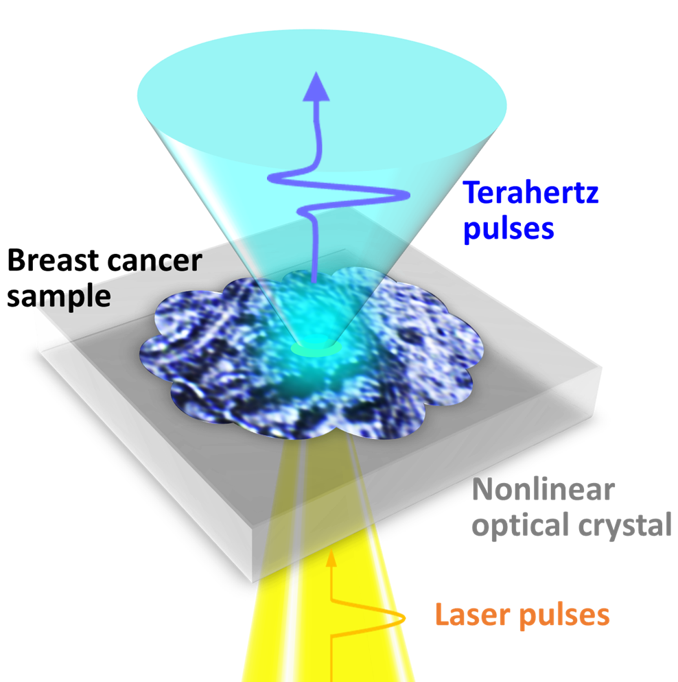
Terahertz imaging
can distinguish cancer tissue from normal tissue without staining and radiation exposure.
However, it was still difficult to identify an individual DCIS lesion (which typically range from 50 to 500 µm) by terahertz imaging due to its diffraction-limited spatial resolution of just several millimeters.
ResOU